Splatting leaves graphics and turns up in the CT scanner
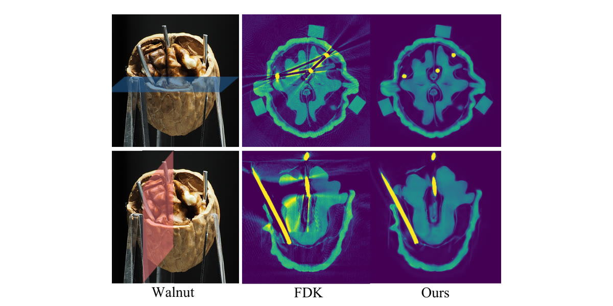
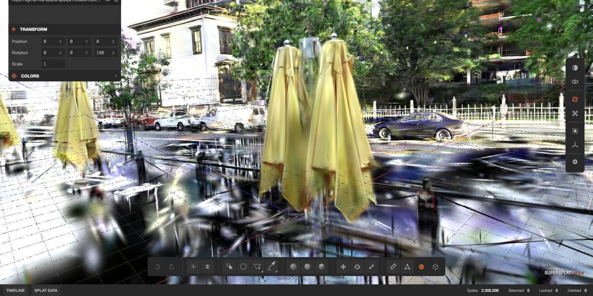
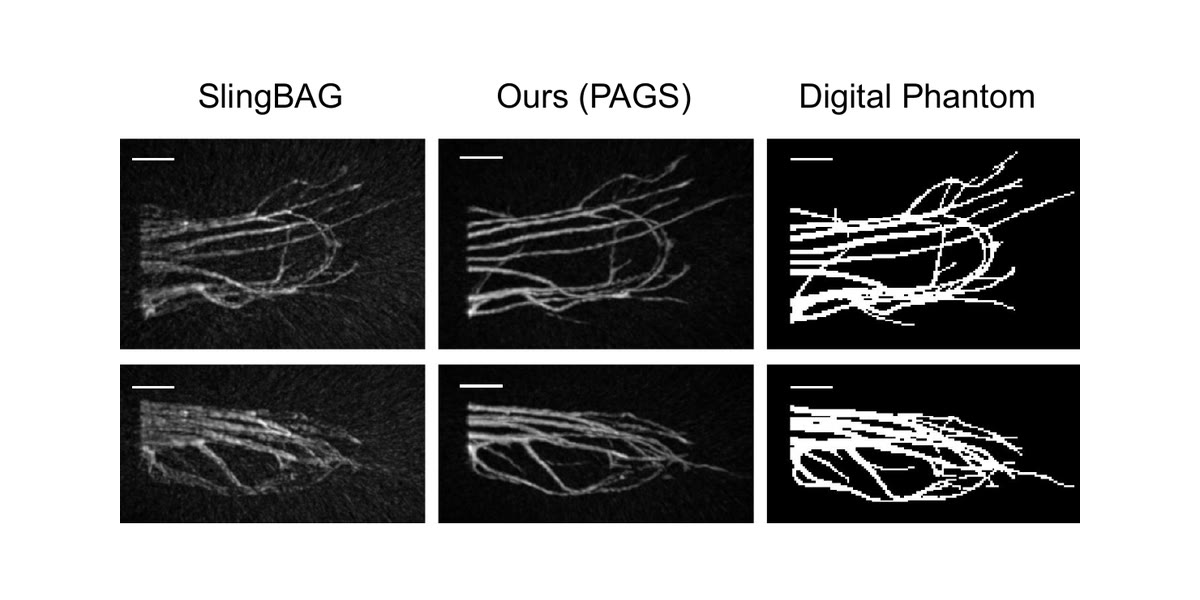
Figure: Choi et al., KAIST — Computer Graphics Forum 45(2) · Research

Aug 14, 2026, 7:15 AM ETResearch
Kiseok Choi, Min H. Kim and colleagues have adapted gaussian splatting into a reconstruction method for cone-beam CT, correcting the streaks and dark bands that metal implants throw across a scan by modeling the polychromatic X-ray beam that causes them.
Why it matters: Splatting was built to render what a camera saw. This is the same representation running in reverse, as a solver for a physical inverse problem in a medium that has nothing to do with photography.
That generalization is the real story: gaussians are becoming a way to represent volumes, not just a way to draw them.
The problem: Cone-beam CT fires X-rays across a spectrum of energies, but conventional reconstruction pretends the beam is a single energy. Dense material like a dental implant or a surgical screw breaks that assumption badly — the beam hardens as it passes through, and the scan fills with streaks around exactly the anatomy a clinician needs to see.
Neural reconstruction methods improved on this but are, in the authors' framing, too computationally expensive to deploy.
Zoom in:
- The method folds a polychromatic X-ray projection model, material-dependent attenuation profiles, and system response into a gaussian splatting framework.
- It is self-calibrating: reconstruction parameters and the X-ray spectrum itself are optimized jointly during training.
- It needs no manual metal masks and no strong prior assumptions — the segmentation step earlier methods depended on simply goes away.
- The team also released a synthetic CBCT data pipeline, validated against a Monte-Carlo X-ray simulator, plus datasets with severe metal artifacts.
What's next: The authors report beating state-of-the-art artifact suppression on both synthetic and real scans. The claim to watch is efficiency: if a splat-based reconstruction is genuinely cheap enough to run on scanner hardware, it lands somewhere neural methods never reached.